
Figure 3.
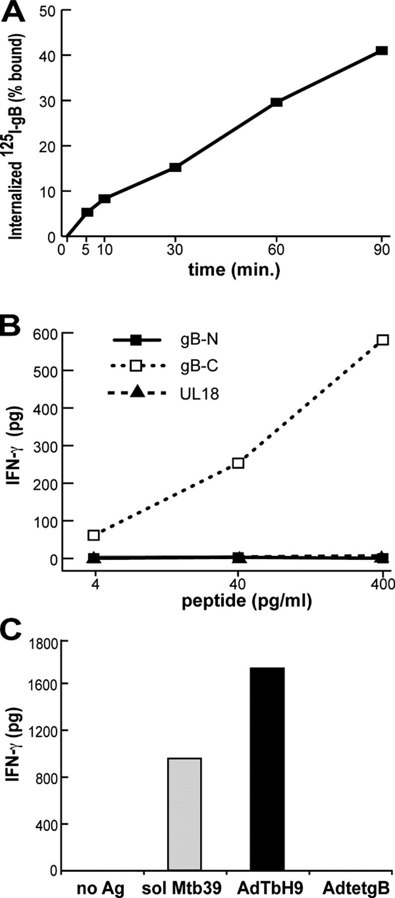
Glial cells internalize soluble gB, and present gB peptides and a soluble TB antigen. (A) His16 cells were incubated with 125I-gB at 4°C, washed, warmed to 37°C, and the cell surface gB was removed with citrate buffer before counting cell-associated (internalized) 125I. Background (cells not warmed to 37°C) was subtracted from each value. (B) His16 cells were incubated with pools of peptides (15 mers overlapping by 10 residues) making up the NH2-terminal (gB-N, residues 1–440) or the COOH-terminal (gB-C, residues 430–907) half of gB or all of UL18, for 6 h before incubation with gB10/G3 CD4+ T cells for 24 h. (C) His16 cells were incubated with medium alone (no Ag) or with 1 μg/ml of soluble mtb39, and TbH9-9 (mtb39-specific) T cells, or were infected with 100 PFUs/cell of AdTbH9 or AdtetgB for 24 h before addition of TbH9-9 T cells. IFN-γ was measured in B and C.