
Figure 1.
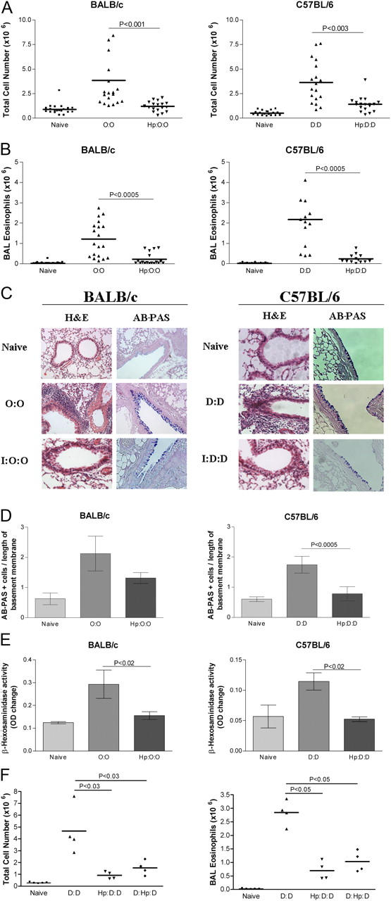
Airway cell infiltration and tissue pathology are inhibited in H. polygyrus-infected mice. BALB/c or C57BL/6 mice were sensitized to OVA or Der p 1, respectively, by two i.p. injections of 10 μg of allergen adsorbed to Alum; 14 and 17 d after the second sensitization, mice were given soluble allergen by direct tracheal inoculation. Lavage cells were recovered 24 h after final airway challenge (day 31), and histological sections made of lung tissues. O:O denotes OVA sensitized (day 0 and 14), and challenged (day 28 and 30). D:D denotes Der p 1 sensitized (day 0 and 14), and challenged (day 28 and 30). Cells were harvested at day 31. Hp:O:O and Hp:D:D are mice infected with H. polygyrus 28 d before allergen sensitization; D:Hp:D are mice infected 14 d after sensitization, but before challenge. (A) Total lavage cell numbers. Data represent means and individual data from four experiments (18–20 mice per group) using the same protocol. Horizontal bars denote paired experimental groups for which statistical significance is displayed in the figure. (B) Eosinophil numbers. All four experiments displayed significant reductions in airway eosinophilia in infected-allergen challenged animals compared with uninfected allergen challenged littermates. (C) Formalin-fixed lung 5-μm sections from naive, allergic, and infected-allergic mice stained with hematoxylin and eosin (H&E) for nuclear staining of infiltrating cells or stained with AB-PAS for mucin-producing goblet cell numbers. (D) Enumeration of goblet cells, stained with AB-PAS in the three groups of mice. Data represent mean and SEM of five mice, with a mean score from three representative lung sections per mouse. (E) β-Hexosaminidase in BALF of allergic and infected mice. (F) Total cells and eosinophils in mice infected before or after allergen sensitization.