
Figure 5.
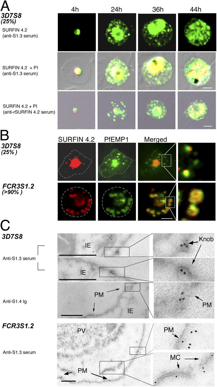
SURFINs expressed in 3D7S8 and FCR3S1.2 are cotransported with PfEMP1 to the host PM. (A) Indirect immunofluorescence assay (IFA) on air-dried monolayers of 3D7S8 IEs at various stages of maturation. (top) Conventional IFA with SURFIN4.2-directed anti-S1.3 antibodies at 4, 24, 36, and 44 h a.i.; (middle and bottom) merged double-stained confocal IFA of PI-stained parasite nuclei (red) with Alexa 488–labeled SURFIN4.2 (green). SURFIN was detected with either anti-S1.3 (middle) or anti-rSURFIN4.2 antibodies (bottom); yellow, label overlay. Both antibodies detected SURFIN4.2 in ∼25% of examined parasites at ∼24 h a.i. in dot-like transport vesicles outside the parasitophorous vacuole. (B) Dual localization of TRITC-labeled SURFIN4.2 (red) and Alexa 488–labeled PfEMP1 (green) showed that SURFIN4.2 and PfEMP1 are colocalized in cytosolic structures of ∼25% 3D7S8 IEs (compare with the merged fluorescence pattern). An expressed SURFIN was detected in >90% of trophozoite-stage FCR3S1.2 IEs (∼24 h a.i.) and colocalized with PfEMP1 in LMVs. Dashed circles indicate the area of the IE. (C) Immunoelectron microscopy studies on 3D7S8 and FCR3S1.2 IEs. SURFIN detected with either anti-S1.3 serum or affinity-purified anti-S1.4 immunoglobulins is present in the PM, as well as in the erythrocyte cytosol associated with Maurer's clefts (MC) as indicated by arrows. Bars: (A and B), 2 μm; (C), 0.5 μm.