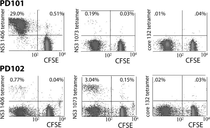
Figure 3.
Proliferation of HCV-specific tetramer+ CD8+ T cells. On day 8 of culture in HCV peptide–stimulated T cell lines derived from PD101 and PD102, CFSE-labeled tetramer+ cells were detected. In brief, 107 PBMCs were cultured in the presence of four HCV epitope peptides (NS3 1406–1415 [KLVALGINAV], NS3 1073–1081 [CINGVWCTV], and core 132–140 [DLMGYIPLV]) with IL-2 (0.5 ng/ml) added on days 0 and 3. Because the CFSE signal is diluted with each cell division, signals in the left upper quadrant represent viral-specific CD8+ T cells that have proliferated in culture. Those in right upper quadrant represent tetramer+ T cells that have not divided (reference 14). (A) In PD101, the NS3 1406–specific CD8+ T cells expanded after peptide stimulation (representing 29% of gated CD8+ T cells) and NS3 1073–specific CD8+ T cells demonstrated modest proliferation, whereas the other epitope tetramer+ cells were viable but failed to expand, as indicated by the tetramer+ cells in the upper right quadrants of the dot plots. Analysis from two other time points after infection revealed similar results. (B) For PD102, in vitro culture for 8 d led to proliferation of NS3 1406– and NS3 1073–specific CDD8+ T cell responses. In contrast, neither core-specific nor NS5B 2594–specific responses expanded in culture. Analysis from two other time points revealed similar results.

