
Figure 1.
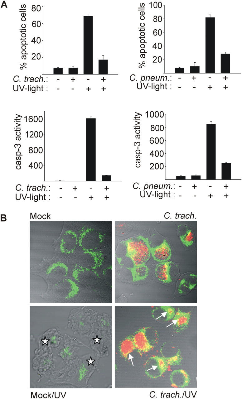
Infection with C. trachomatis or C. pneumoniae inhibits UV light–induced apoptosis. Hep2 cells were infected with Chlamydia or not (mock). After 24 (C. trachomatis) or 48 h (C. pneumoniae), apoptosis was induced by UV irradiation (1,000 J/m2). 6 h later, cells were analyzed. (A) Nuclear apoptosis and caspase 3–like activity. Cells were stained with Hoechst dye and nuclear apoptosis was scored under a fluorescence microscope (top; at least 300 nuclei were counted per sample and values are mean/SD of triplicates) or lysed, and DEVD cleaving activity was measured in cell extracts (bottom; mean/SD of triplicate measurements). (B) Cytochrome c release from mitochondria. Cells were stained for cytochrome c (green) and chlamydial LPS (red), and analyzed by laser-scanning microscopy. Arrows point at chlamydial inclusions in cells that have retained cytochrome c in their mitochondria. Asterisks indicate noninfected cells with typical apoptotic morphology and release of cytochrome c from mitochondria (the cytochrome c signal is diminished upon release). The figure shows results typical of three independent experiments.