Figure 3.
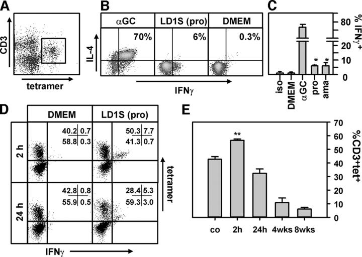
IFNγ production in Leishmania-infected livers. (A) CD1d-tetramer staining. Liver lymphocytes from infected C57BL/6 mice were isolated by Percoll gradient centrifugation and stained with PeRCP-conjugated anti-CD3ɛ mAb and αGC-loaded PE-conjugated CD1d-tetramers. The tetramer positive, CD1d-reactive T cell subset is identified by flow cytometry and shows the characteristic intermediate CD3 expression. (B–E) Intracellular cytokine staining. Groups of C57BL/6 mice were injected intravenously with medium (DMEM), 200 ng αGC, 107 stationary phase LD1S promastigotes (pro), or 107 amastigotes (ama) from infected hamster spleen, and lymphocytes were prepared and stained 2 h after the inoculation (B and C) or at the times indicated (D and E) as described before. Intracellular cytokine staining was performed using the Cytofix/Cytoperm Kit from Becton Dickinson with FITC-conjugated anti-IFNγ and allophycocyanin-conjugated anti–IL-4. The analysis in B and C was gated on CD3(+)tet(+) cells. Staining with FITC-conjugated isotype-specific antibody was performed to control for background (iso). At least three independent triplicate experiments were performed, and one representative experiment is shown. For the analysis in D and E, cells were gated on CD3. One experiment was performed with three to five mice per time point. Asterisks indicate statistically significant differences relative to DMEM-injected mice (*, P < 0.02; **, P < 0.01; unpaired Student's t test).

