
Figure 2.
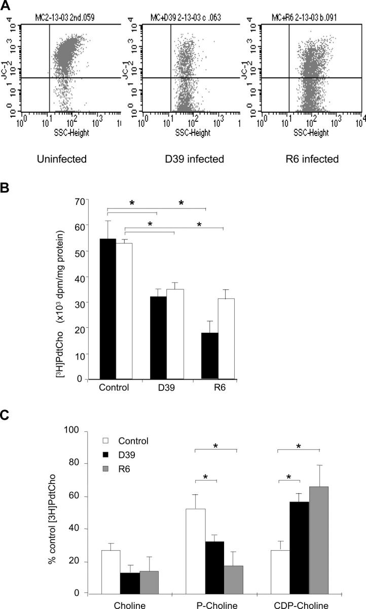
Inhibition of PtdCho biosynthesis by S. pneumoniae induces apoptosis. (A) FACS® analysis of mitochondrial membrane potential of control (left) and infected (middle and right) microglia. Dot plots show side scatter signals (x axis) versus fluorescence intensity (y axis) for microglia cells. Shift of fluorescence of JC-1 from 590 (FL2) to 530 nm (FL1) indicates membrane depolarization. Analysis representative of six independent experiments. (B) Rate of PtdCho biosynthesis. [methyl-3H]choline–labeled human primary microglia (black bars) or cultured cells (white bars) were infected with 2 × 10⁷ cells of strain D39 or R6 pneumococci for 4 h. PtdCho synthesis was measured by scintillation counting and values were normalized to the protein concentration of the sample. Values are mean ± SE of 10 duplicate experiments. *, P < 0.05. (C) Distribution of [methyl-3H]choline incorporation into water-soluble choline intermediates in microglia. *, P < 0.05. Total cpm incorporated were 292,536 ± 68,812. Bacteria alone incorporated 13,410 ± 4,797 (4.5%).