Figure 6.
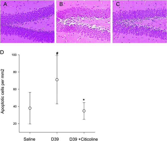
Protection from pneumococcal-induced apoptosis in the hippocampus in vivo. Three groups of six mice were injected via lumbar puncture with saline or pneumococci as indicated. One group also received citicoline intraperitoneally 24 h before infection and via lumbar puncture at the time of infection. Mean bacterial number in cerebrospinal fluid for infected animals was 107 bacteria/ml for pneumococci alone and 3 × 10⁷ bacterial/ml for pneumococci with citicoline. Histopathology of the dentate gyrus: (A) normal control, (B) infected with pneumococcus, and (C) infected with pneumococcus and treated with citicoline. (D) Number of apoptotic neurons per mm2 in the hippocampus ascertained at 24 h of infection. The data were statistically analyzed by a One Way Analysis of Variance (P < 0.05) followed by a Student-Newman-Keuls test. #, the difference between controls and meningitis (P = 0.01). *, the difference between untreated meningitis and citicoline treatment (P = 0.02).

