Figure 4.
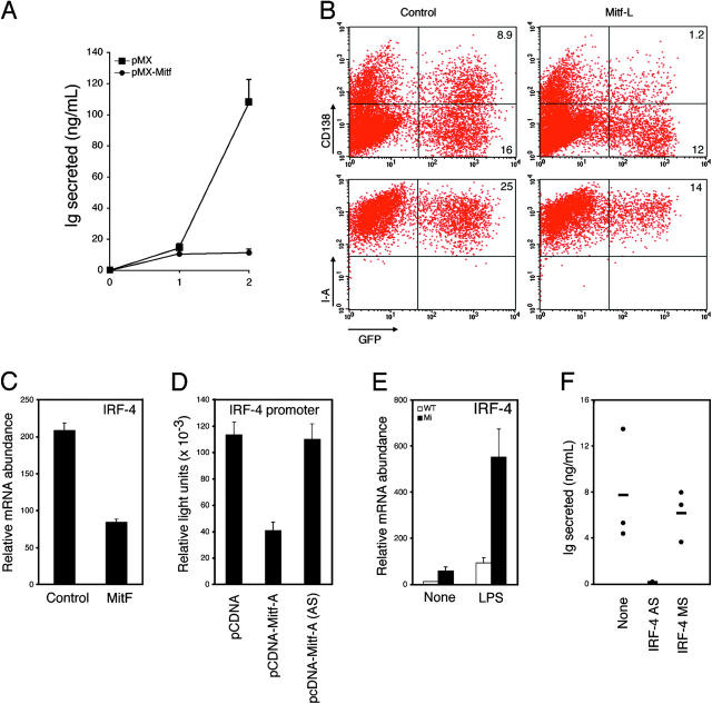
Mitf represses plasmacytoid differentiation via IRF-4. (A) B cells from wild-type C57BL/6 animals were stimulated with 25 μg/ml LPS for 24 h and infected with a control (pMX) or Mitf-A–expressing (pMX-Mitf) GFP retrovirus in the continued presence of LPS. (A) On day 2 after infection, GFP-positive cells were sorted and returned to medium containing LPS, and IgM secretion was assessed 1 and 2 d thereafter by ELISA. (B) On day 2 after infection, cells were assessed for syndecan-1 (CD138) and MHC class II (I-A) expression by flow cytometry. (C) GFP-sorted B cells infected with a control or Mitf-expressing retrovirus, as in A, were returned to medium containing LPS for an additional 24 h and assessed for the expression of IRF-4 by real-time PCR. (D) The ability of Mitf to regulate the IRF-4 promoter was assessed by transient transfection in M12 B cell lymphoma cells, using a control, Mitf-A, or Mitf-A antisense (AS) expression construct. Firefly light units were normalized against control Renilla luciferase activity. (E) Naive B cells from wild-type or mi Rag-deficient chimeras were incubated in vitro in the presence or absence of LPS for 24 h. The expression of IRF-4 was assessed by real-time PCR. (F) Naive B cells from mi Rag-deficient chimeras were incubated in vitro without mitogenic stimulation in the presence or absence of IRF-4 antisense (AS) or missense (MS) oligonucleotides. After 10 d, supernatants were assessed for IgM secretion by ELISA. Standard deviations reflect triplicate samples performed simultaneously, and are representative of at least three experiments.

