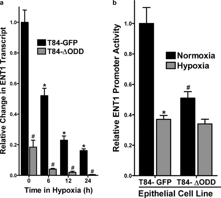
Figure 7.
Functional regulation of ENT1 expression in epithelial cells expressing oxygen-stable HIF-1α. (a) T84-GFP and T84-ΔODD cells were exposed to indicated periods of hypoxia and examined for ENT1 expression by real-time PCR. Data were calculated relative to β-actin and are expressed as relative change ± SD at each indicated time. Results are derived from three experiments in each condition (*P < 0.01, different from normoxia and ENT2; #P < 0.01, different from normoxia). (b) T84-GFP and T84-ΔODD cells were transiently transfected with the wild-type ENT promoter construct (ENT1-756) and exposed to hypoxia or normoxia (24 h). Relative activity was assessed by luciferase relative to empty vector (pGL3). Results are presented as relative change in activity above PGL3 background (relative to control plasmids expressing Renilla in each condition).

