Figure 1.
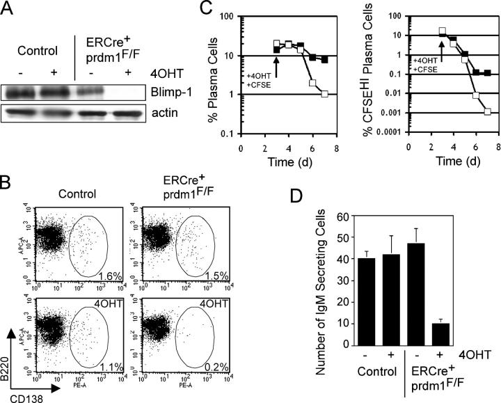
Loss of Blimp-1 in vitro. Splenocytes were treated with LPS for 3 d in vitro followed by 4 d of 4-hydroxytamoxifen (4OHT) or sham treatment. (A) Western blot for Blimp-1 and actin protein in ERCre + prdm1 F/F and control cultures treated with 4OHT and sham. (B) CD138 vs. B220 staining of splenocyte cultures. (left) Control; (right) ERCre + prdm1 F/F cultures; (top) sham; (bottom) 4OHT treated. The frequency of gated CD138HI plasma cells is indicated. Depicted results are representative of five experiments. (C) Kinetics of plasma cell disappearance after 4OHT treatment. (left) The percent of total B220LOCD138HI plasma cells in control (solid square) and ERCre + prdm1 F/F (open square) cultures for 4 d after 4OHT treatment. (right) The percent of CFSEHI plasma cells in these cultures. (D) IgM-secreting cells as assessed by ELISPOT analysis for ERCre + prdm1 F/F and control cultures treated with 4OHT and sham. The y axis represents number of IgM-secreting cells per 2,000 cells. C and D are representative of two experiments.

