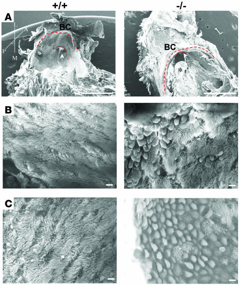
Figure 6. Scanning electron micrographs of middle ear cavities from wild-type (+/+) and Eya4–/– (–/–) mice.
(A) Lateral view of the eustachian tube ostium (arrow) from 16-month-old mice. The ostium is malpositioned in the Eya4–/– ear, near to the edge of the middle ear bony capsule (BC). The junction of the bony capsule with the temporal bone (thick dashed line) and the osseous portion of eustachian tube (thin dashed line) are highlighted. A polyp (asterisk) is obstructing the Eya4–/– OpTC. Axis: A, anterior; P, posterior; L, lateral; M, medial. Scale bars: 1 mm. (B) Lateral view of eustachian tube ostia epithelia in 3-week-old mice shows increased numbers of goblet cells and swollen epithelia in Eya4–/– mice, although cilia are morphologically indistinguishable from cilia in wild-type mice. Scale bars: 5 μm. (C) The mucociliary epithelia at the eustachian tube ostia of 16-month-old mice show rarefaction of cilia in the mucosa of Eya4–/– mice. Scale bars: 5 μm.

