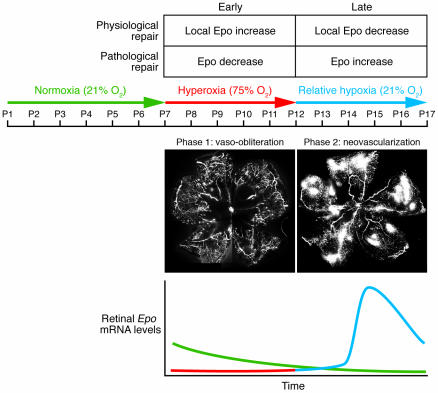
Figure 1. Mouse model of proliferative OIR.
In this model, 7-day-old mouse pups with partially developed retinal vasculature are subjected for 5 days to hyperoxia (75% oxygen), which stops retinal vessel growth and causes significant vaso-obliteration (phase 1). On postnatal day 12, pups are returned to room air, and by postnatal day 17, a florid compensatory retinal neovascularization occurs (shown in white) (phase 2). In the line graph, a representation of retinal Epo mRNA levels in the OIR model during normoxic conditions (green), phase 1 (red), and phase 2 (blue) are shown. As Chen et al. (5) show, in the OIR model during hyperoxia (phase 1), Epo levels are reduced, resulting in pathological elevations of Epo at the late stage of disease development. With Epo treatment during hyperoxia, retinal vasculature is protected and BM-derived EPCs come into the developing vasculature, promoting healthy vessels rather than pathological neovascularization as in the OIR model. Original magnification, ×5.

