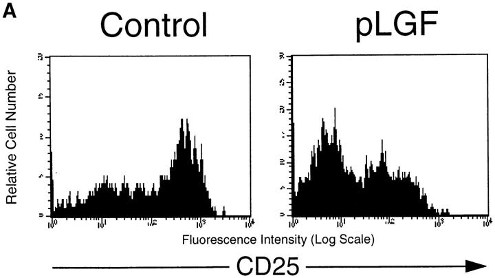
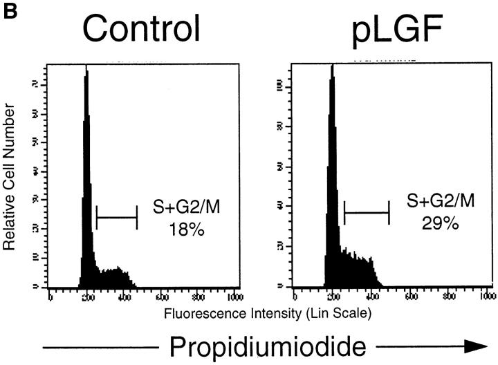
Figure 1.
Transgenic expression of activated p56lck enhances differentiation and proliferation of pre-T cells. (A) CD25 expression profiles of CD44neg TN thymocytes in normal control mice and pLGF transgenic mice expressing active p56lck. Single cell suspensions of total thymocytes from normal control and pLGF transgenic mice were prepared and stained with mAb against a panel of mature lineage markers including CD4, CD8, CD3, B220, NK, Gr-1, Mac-1 (all biotinylated), CD44 (biotinylated), and CD25 (FITC). Cells negative for all lineage markers and CD44 were gated and examined for their CD25 expression profile. (B) Transgenic expression of active p56lck leads to increased levels of CD44neg TN thymocytes in S+G2/M phase of the cell cycle. Total thymocytes from normal and pLGF transgenic mice were prepared and stained with mAb against CD44 (biotinylated) and a panel of mature lineage markers including CD4, CD8, CD3, B220, NK, Gr-1, and Mac-1 (all biotinylated), fixed in 70% ethanol, and stained with propidium iodide (PI). Cells lacking expression of CD44 and all lineage markers were gated and then examined for their DNA profile.


