
Figure 1.
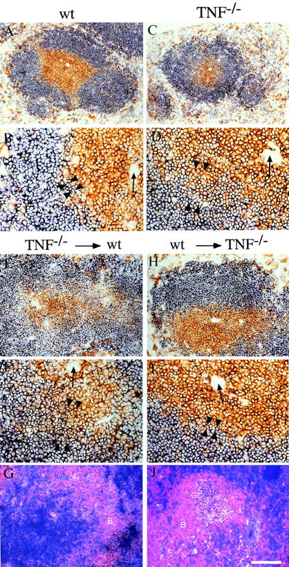
WT bone marrow restores correct follicular structure and GC reaction in lethally irradiated TNF−/− recipients. (A–D) Spleen sections from unmanipulated 6–10-wk-old animals were stained for a combination of CD4+ and CD8+ T cells (brown) and B220+ B cells (blue). (E–J) TNF−/− bone marrow was injected into lethally irradiated WT recipients (E–G) and WT bone marrow into TNF−/− recipients (H–J). 30 wk later, spleen sections were stained by immunohistochemistry for CD4+ and CD8+ T cells (brown) and B220+ B cells (blue) (E, F, H, and I), or sections obtained 8 d after immunization with SRBC were stained for B cells (red) and PNA+ GCs (blue/ purple) (G and J). Arrows, Central arterioles; arrowheads, T–B interface. Bar = 400 μm (A and C), 200 μm (E, G, H, and J), or 100 μm (B, D, F, and I).