
Figure 7.
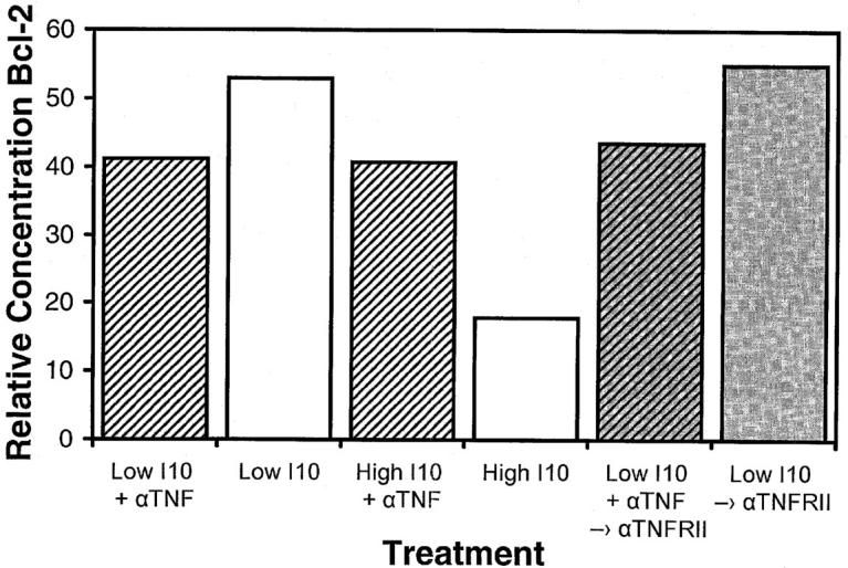
Signaling through TNF-RII is not sufficient to decrease Bcl-2 levels, but TNF-α signaling contributes to this effect. High avidity CTLs were stimulated by transfer to soluble Dd coated wells pulsed with optimal (0.0005 μM) or supraoptimal (50 μM) I10 peptide in either the presence or absence of anti–TNF-α. After 6 h, cells were transferred to either untreated wells or wells coated with anti–TNF-RII and incubated for a further 22–24 h under their initial media conditions. CTLs were then harvested and solubilized with 0.5% Triton X-100 lysis buffer containing protease inhibitors. Lysates were analyzed by Western blot analysis for determination of Bcl-2 concentrations. Actin was used as a control to ensure equivalent loading among the samples. Anti–TNF-α blocked the decrease in Bcl-2 that occurred after high avidity CTLs were stimulated with high peptide–MHC determinant density. Substrate-bound anti–TNF-RII caused no decrease in the levels of Bcl-2, either in the presence or in the absence of anti– TNF-α, indicating that these conditions were not equivalent to stimulation through the TCR by supraoptimal levels of peptide–MHC.