
Figure 8.
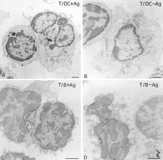
Analysis by electron microscopy of conjugates between T cells and either DCs with (A) or without (B) Ag, or immunogold-stained B cells with (C) or without (D) Ag. For each image, the T cell engaged in the conjugate is at the left of the APC. Bars, 1 μm.