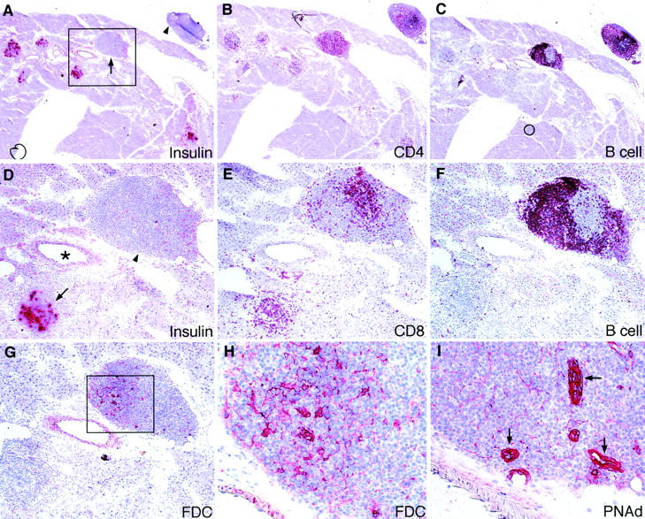
Figure 4.

Organization of newly formed lymphoid tissues in RIP-GP mice after repetitive priming with H8-DC. Pancreata were analyzed by immunohistochemistry on day 21 by staining for the indicated markers. (A) Newly formed organized lymphoid tissue was found periductally in the pancreatic parenchyma (arrow); a pancreatic lymph node located outside the pancreatic parenchyma is also shown (arrowhead). Distribution of infiltrating (B) CD4+ T cells and (C) B cells in islets and newly formed lymphoid tissues. (D) Magnification of the boxed area in A showing an insulin-positive islet (arrow) and periductal, de novo–formed lymphoid tissue (arrowhead) in close vicinity to an artery (*). Distinct spatial distribution of (E) CD8+ T cells, (F) B cells, and (G) follicular DC (FDC) in the newly formed lymphoid tissue. (H) Magnification of the boxed area in G showing the 4C11+ follicular DC network. (I) Staining for PNAd. The region corresponding to the boxed area in G was photographed showing PNAd+ blood vessels in the newly formed lymphoid tissue. Original magnifications: A–C, ×24; D–G, ×63; H and I, ×250.
