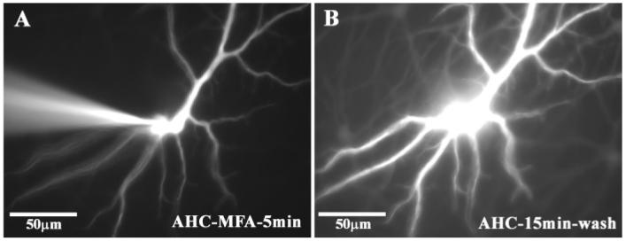
Fig. 4.
Reversal of Lucifer Yellow coupling in real time after the block of gap junctions with meclofenamic acid. (A) shows the electrode impaling a single A-type HC after 5 min of dye injection with 4% Lucifer Yellow during perfusion with 100 μM MFA. (B) The electrode was withdrawn and after a 15 min wash, Lucifer Yellow spread to other A-type HCs. Same field as A, as shown by identical morphology of filled cell. This indicates that coupling was reversible after the washout of meclofenamic acid.

