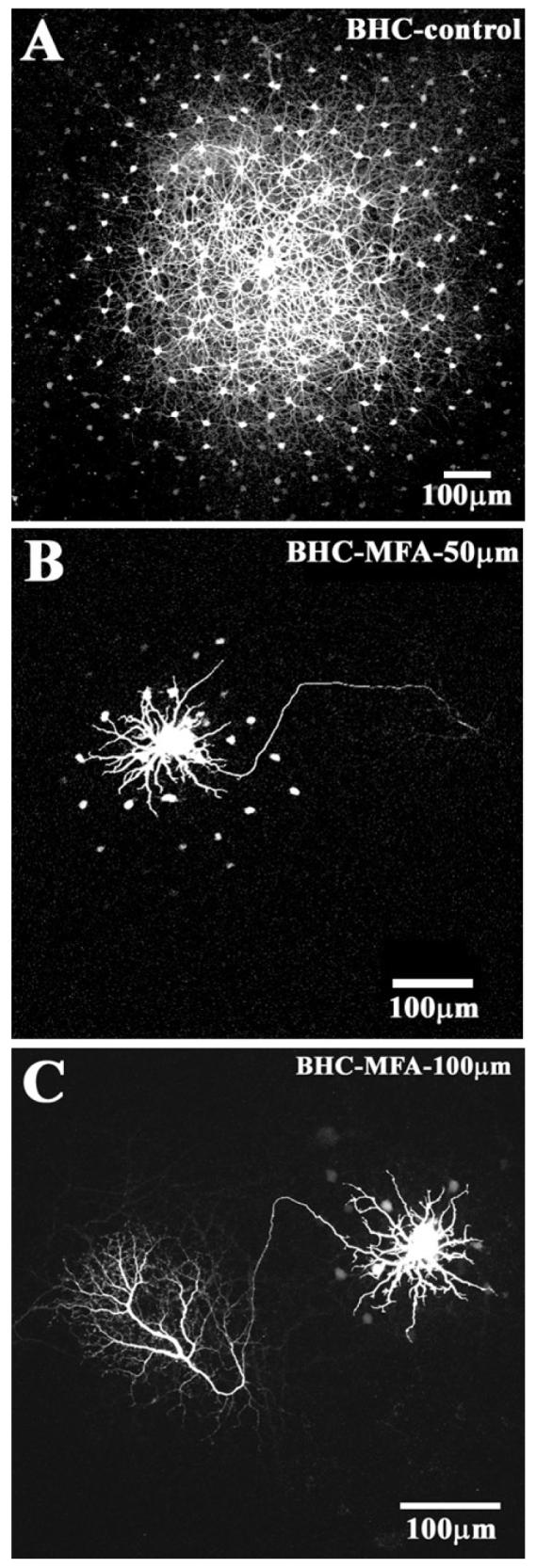
Fig. 5.

The block of gap junction coupling in B-type HCs. (A) Control experiment shows a large patch of coupled B-type HCs after 10 min of dye injection followed by 10 min of diffusion. (B) The same protocol in the presence of 50 μM MFA produced a dramatic reduction in coupling. A single prominent B-type HC was surrounded by a few coupled somas and the axon was brightly stained. (C) Increasing the dose of MFA to 100 μM further reduced the coupling of B-type HCs and produced axon and axonal terminal.
