Figure 2.
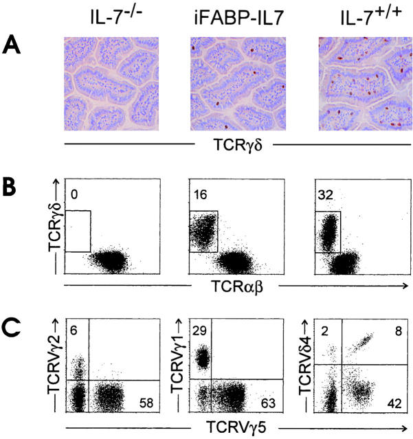
Restoration of typical TCR-γδ IELs in iFABP-IL7 mice. (A) Frozen sections of small intestinal villi from IL-7−/− (left), iFABP-IL7 (center), and IL-7+/+ (right) mice were stained with mAb specific for TCR-γδ as described in Materials and Methods. Note the complete absence of TCR-γδ cells from IL-7−/− mice and partial restoration of TCR-γδ cells to iFABP-IL7 mice. (B) Small intestinal IELs were isolated from IL-7−/−, iFABP-IL7, or age-matched IL-7+/+ control animals. The cells were stained with mAb directed against CD3ε, TCR-αβ, and TCR-γδ and then analyzed by fluorescence flow cytometry. CD3ε+ cells within the forward versus side scatter lymphocyte gate were analyzed for TCR expression. The numbers indicate the percentage of TCR-γδ+ cells among total CD3ε+ cells. (C) Small intestinal IELs from iFABP-IL7 mice were stained with mAb directed against either TCR-γδ, TCRVγ5, and TCRVγ2 or TCRVγ1 or CD3ε, TCR-αβ, TCRVγ5, and TCRVδ4. The numbers reflect the percentage of the indicated TCRVγ+ cells among total TCR-γδ cells. Since antibodies directed against all TCR-γδ and TCRVδ4 compete for binding, CD3ε+TCRαβ− IELs were positively gated and analyzed for expression of TCRVγ5 and TCRVδ4.

