
Figure 7.
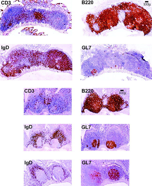
Visualization of germinal centers in iFABP-IL7 Peyer's patches. Peyer's patches were dissected from the small intestine of age-matched control IL-7+/+ mice (top) or iFABP-IL7 mice (bottom). 6-μm frozen sections were stained as described in Materials and Methods with mAbs that identified total B cells (B220+), total T cells (CD3+), naive B cells (IgD+), or germinal center B cells (GL7+). The iFABP transgene restored germinal centers to approximately the size visualized in normal mice, despite the concurrent deficiency in naive B cells.