
Figure 4.
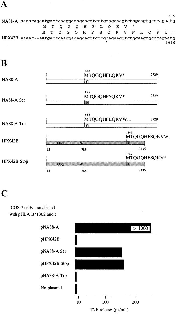
Identification of differences between HPX42B and NA88-A cDNAs important for their differential ability to generate a signal stimulating TNF release by M88.7 T cells. (A) Comparison of NA88-A and HPX42B nucleotide and encoded protein sequences in the region coding for antigenic peptide. Potential initiation and stop codons are indicated in bold characters. (B) Schematic representations of normal and mutated NA88-A and HPX42B cDNA sequences. Boxes marked P code for antigenic peptides and are white for the NA88-A peptide and grey for the HPX42B peptide (as marked above each box). For NA88-A Ser, a Leu codon, CTG, has been changed to a Ser codon, TCG, within the peptide-coding region. For NA88-A Trp, a stop codon tag immediately following the antigenic peptide's terminal Val codon has been changed to a Trp codon, tgg. For HPX42B Stop, a Trp codon, tgg, immediately following a Val codon has been changed to a stop codon, tag. (C) pcDNA3-based plasmids containing cDNAs as described in B were cotransfected into COS-7 cells with pHLA B*1302. The ability of transfected cells to induce TNF release by M88.7 T cells was measured. Note that all results shown here are from transfections carried out simultaneously in parallel, allowing comparison of results between samples. TNF release values for pNA88-A are higher than those shown in the separate experiment of Fig. 1 and Fig. 3, as results depend on COS-7 cell transfection efficiencies and the condition of the WEHI cells used for TNF assays, both of which vary between individual experiments.