
Figure 2.
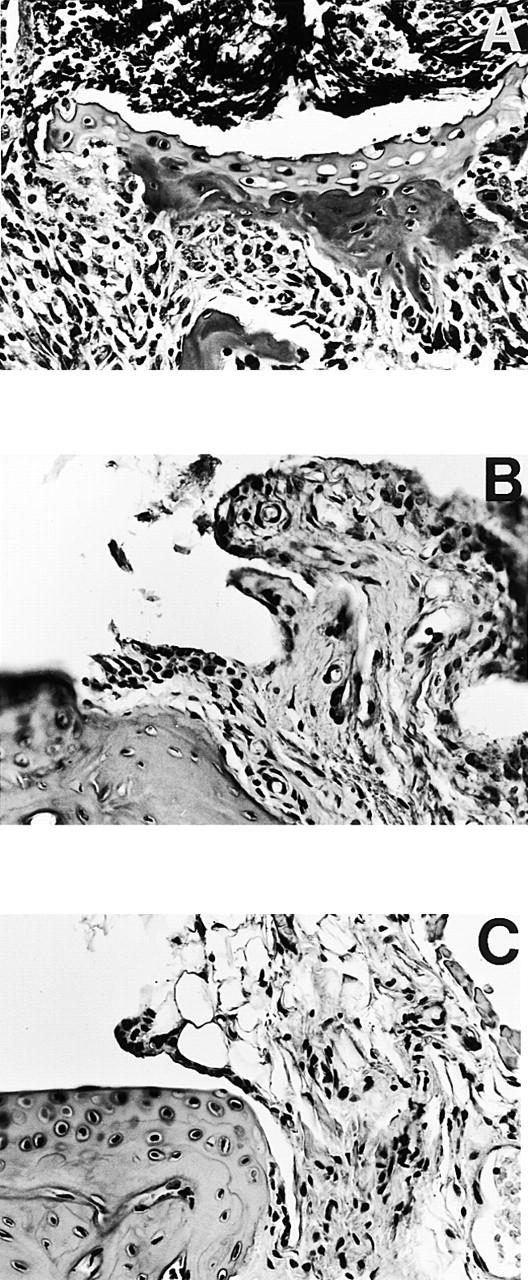
Histopathology of tarsal joints from FcRγ+/+ and FcRγ−/− DBA/1 mice 80 d after CII immunization. Severe arthritis was seen in FcRγ+/+ mice (A) with inflammatory cellular infiltrate, invasive pannus, and erosions of cartilage and bone clearly detectable. The few FcRγ−/− mice that developed disease (B) showed proliferation of synovial lining layer, synovial villi formation, but absence of cellular infiltrate and erosions. Joints of nonaffected FcRγ−/− mice (C) were normal in appearance, with normal synovia and smooth intact cartilage. Representative sagittal paraffin sections with hematoxylin-eosin stain; original magnifications: (A) ×20; (B and C) ×50.