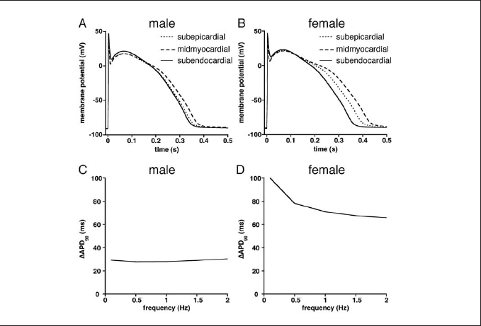
Figure 2. (A,B) Superimposed action potentials (0.5 Hz) of subepicardial, midmyocardial, and subendocardial model cells of male (A) and female (B). (C,D) Differences between longest (midmyocardial) and shortest (subendocardial) action potentials (ΔAPD90) at various stimulus frequencies in male (C) and female (D) model cells.
