
Figure 1.
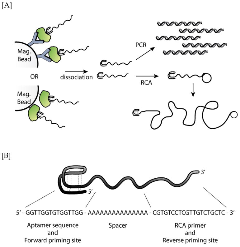
[A] Schematic of the aptamer binding biodetection process. Briefly, either anti-protein antibody or biotinylated protein target was first immobilized onto magnetic beads. After incubation with the DNA aptamers, unbound aptamers were removed. The target-bound aptamer constructs were dissociated and the aptamers were analyzed in real time via either PCR or RCA. [B] Sequence of the 50nt ssDNA aptamer construct, comprised of a thrombin recognition domain, a poly-A linker spacer, and a short RCA primer.