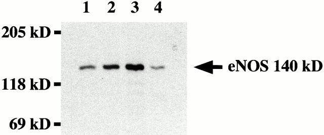
Figure 3.
eNOS expression in rat aorta. Homogenates of aortic endothelium from young (lane 1), middle-aged (lane 2), and old (lane 3) rats were separated by SDS-PAGE and analyzed by Western blotting for eNOS expression. The position of the molecular mass markers is indicated (expressed in kD). Human umbilical vein endothelial cells (lane 4) were used as positive control.

