Figure 1.
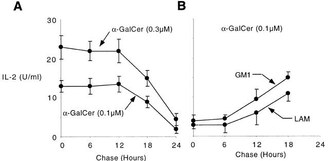
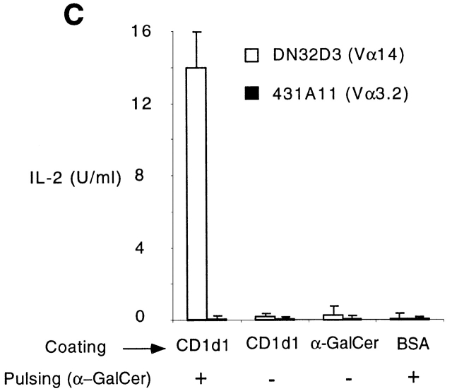
Long life span of CD1d1–glycolipid complexes. (A) Plate-bound CD1d1 (2.5 μg/ml) was loaded with 0.1 or 0.3 μM αGalCer for 2 h, washed, and chased for different periods of time before adding the Vα14-Jα281 NKT cell hybridoma DN32D3 and measuring IL-2 release in the supernatant (mean ± SD). (B) Plate-bound CD1d1 was first incubated with 30 μg/ml of LAM or GM1 for 6 h, washed, and chased for different periods of time. 0.1 μM αGalCer was added for 2 h and washed before incubation with DN32D3. (C) Microwells were coated overnight with 2.5 μg/ml CD1d1 or BSA or 0.1μM αGalCer, washed, then pulsed for 2 h with 0.1 μM αGalCer as indicated, and washed again before adding the CD1d-restricted Vα14 hybridomas DN32D3 or a control CD1d-restricted non-Vα14 hybridoma, 431.A11 (Vα3.2-Jα8/Vβ8), and measuring IL-2 release (mean ± SD). 431A11 and DN32D3 produced similar amounts of IL-2 when stimulated with CD1d1-expressing thymocytes.


