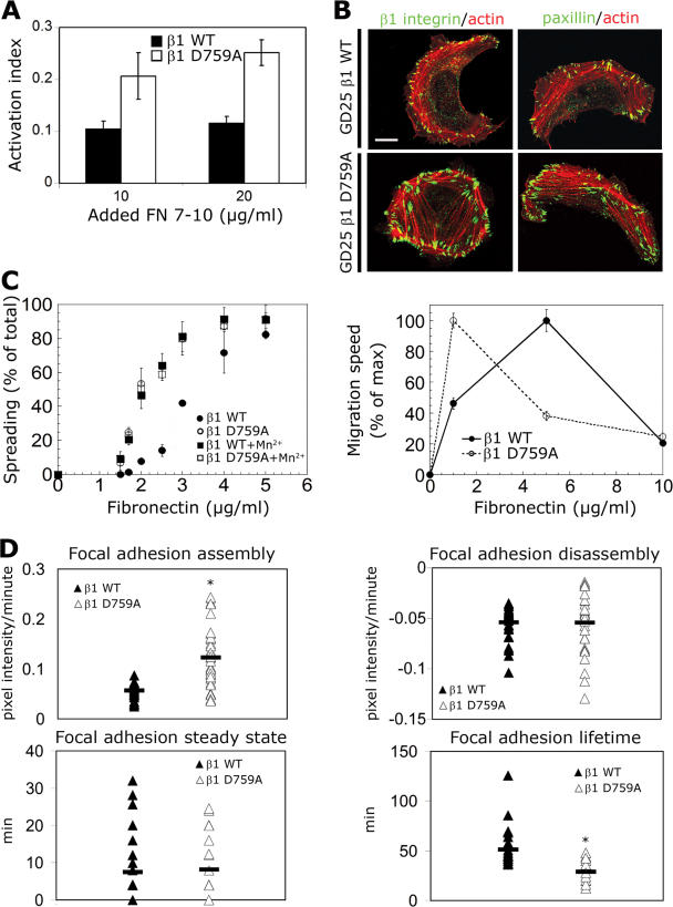
Figure 7.
Activated β1 integrin increases FA turnover and promotes cell migration and spreading at low matrix density. The human WT β1-integrin chains or activated mutant (D759A) were stably expressed in β1 integrin–deficient GD25 cells, and their adhesive properties were analyzed. (A) The integrin affinity state for both cell types was measured with the FNIII7-10 binding assay as described in Fig. 6 A. (B) Confocal images of GD25/WT and D759A β1 cells spread on 10 μg/ml FN and immunostained to visualize β1 integrin, paxillin, and filamentous actin. β1 integrin was labeled with the 4B7R mAb, which recognizes the human β1-integrin chain. (C) Cell spreading and migration assays of GD25/WT and D759A β1 cells. Spreading of both cells types with or without 0.5 mM MnCl2 treatment was measured as described in Fig. 6 B. Migration of both cells types was analyzed using time-lapse phase-contrast video microscopy and cell tracking as described in Fig. 2 B. (D) GD25 cells stably expressing EGFP-zyxin and either WT β1 or D759A integrin were spread on 5 μg/ml FN and monitored by time-lapse video microscopy at 4-min intervals for 6 h. FA dynamics were assessed as described in Fig. 4. Four parameters for FA located at the leading edge of migrating cells were measured: assembly and disassembly speed, steady-state duration, and lifetime. Horizontal bars are the mean of all FAs. Error bars indicate SD. *, P < 105. Bar, 20 μm.

