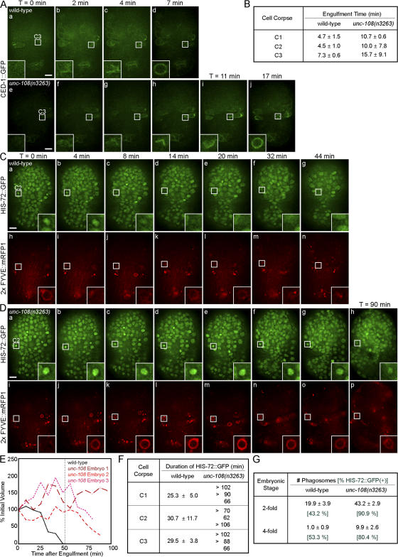
Figure 5.
unc-108(n3263) causes defects in the engulfment and degradation of apoptotic cells. Time-lapse images of embryos expressing Pced-1 ced-1::gfp (A) or coexpressing Phis-72 his-72::gfp and Pced-12× FYVE::mrfp1 (C and D). Insets are a 3× view of the boxed areas. The anterior is shown on top and the ventral side faces outward. (A) Engulfment of cell corpse C3 visualized by the clustering CED-1::GFP on extending pseudopods. Time (T) = 0 min, the time point immediately before budding pseudopods are detected. The enclosure of the CED-1::GFP circle marks the completion of engulfment. (B) Time required for the engulfment of C1, C2, and C3 in wild-type and unc-108(n3263) embryos given as mean ± SD (n = 3). (C and D) The degradation of C2 cell corpses. Nuclei of all cells are marked with HIS-72::GFP and phagosomal membranes are marked with 2× FYVE::mRFP1. T = 0 min, the completion of engulfment marked by recruitment of 2× FYVE::mRFP1. Bars, 5 μm. (E) Relative volumes of representative phagosomes containing C1 plotted against time. (F) Duration of apoptotic cell HIS-72::GFP in phagosomes. For wild-type samples, results are listed as mean ± SD (n = 3). Results for three individual cell corpses are reported for unc-108(n3263). (G) The number of phagosomes observed in twofold stage embryos reported as mean ± SD (n = 15). Numbers in green represent the percentage of phagosomes containing HIS-72::GFP(+) cell corpses.

