Figure 6.
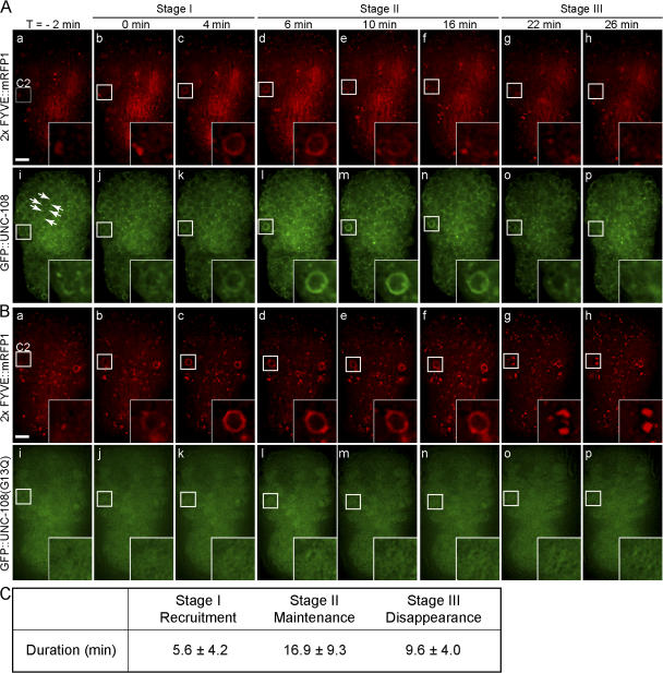
UNC-108 is transiently enriched on phagosomal surfaces. Time-lapse images of wild-type embryos coexpressing Phsp gfp::unc-108 and Pced-12× FYVE ::mrfp1 (A) or Phsp gfp::unc-108(G13Q) and Pced-12x FYVE::mrfp1 (B). Insets are a 3× view of the boxed regions. The anterior is shown on top and the ventral side faces outward. Time (T) = 0 min, formation of a 2× FYVE::mRFP1 circle. (A) Transient enrichment of GFP::UNC-108 to phagosome membranes (i–p). GFP::UNC-108(+) puncta are indicated by arrows. (B) Diffuse cytoplasmic localization of GFP::UNC-108(G13Q) and the failure in recruitment to phagosomes (i–p). Bars, 5 μm. (C) Dynamic pattern of GFP::UNC-108 enrichment on phagosome surfaces in wild-type embryos. See text for the definition of the three stages. Results are reported as mean ± SD (n = 9).

