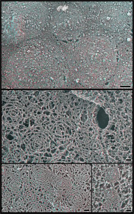
Figure 3.
hSnf7 filaments on the top surface of the cell. (top) Patterns created by hSnf7-1 filaments on the outer surface of whole cells. Shown is the top surface of a COS-7 cell transfected with FLAG hSnf7-1, fixed, and replicated without disruption. Note the subtle circular patterning of particles within the membrane. (middle) Views of hSnf7-1 filaments in the subplasmalemmal “membrane skeleton” revealed by extracting fixed whole cells with detergent. The cell on the left expresses FLAG hSnf7-1, whereas the one on the right does not. (bottom left) Fixed and extracted cell expressing higher levels of FLAG hSnf7-1. (bottom right) Fixed and extracted cell expressing hSnf7-1–mGFP. Bars, 100 nm.

