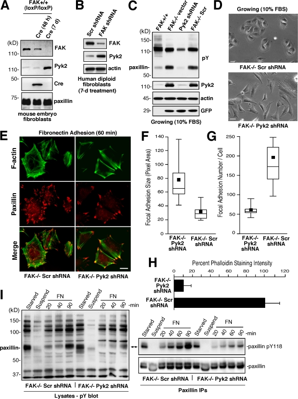
Figure 1.
Pyk2 knockdown affects FAK−/− MEF morphology, FA formation, and paxillin tyrosine phosphorylation. (A and B) Compensatory increase in Pyk2 levels upon inactivation of FAK expression. Lysates from FAK+/+, FAK−/−, and FAK+/+(loxP/loxP) MEFs were analyzed by anti-FAK, Pyk2, Cre recombinase, and paxilllin blotting. Lysates from Ad-Cre–infected FAK+/+(loxP/loxP) MEFs were analyzed after 48 h or 7 d. Paxillin levels were used as a loading control. (B) Human diploid fibroblasts were treated with lentiviral Scr or FAK shRNA and protein lysates were evaluated by anti-FAK, Pyk2, and actin blotting after 7 d. (C) Stable Pyk2 reduction in FAK−/− MEFs by lentiviral anti-Pyk2 shRNA expression. Lysates from the indicated MEFs were analyzed by antiphosphotyrosine (pY), Pyk2, actin, and GFP blotting. (D) Phase-contrast images show that FAK−/− Pyk2 shRNA MEFs exhibit more of a fibroblast phenotype than FAK−/− Scr shRNA control MEFs. Bars, 30 μm. (E) F-actin was reduced and fewer FAs formed in FN-replated (60 min) FAK−/− Pyk2 shRNA compared with FAK−/− Scr shRNA MEFs as determined by phalloidin and anti-paxillin costaining, respectively. Alexa Fluor 350 phalloidin stain is pseudocolored green. Bar, 20 μm. (F) Increased FAK−/− FA size upon reduced Pyk2 expression. Box-and-whisker plots of paxillin-stained area within FAK−/− Scr shRNA or FAK−/− Pyk2 shRNA MEFs plated on FN for 60 min (n = 15 cells per point). Box-and-whisker diagrams show the distribution of the data: square, mean; bottom line, 25th percentile; middle line, median; top line, 75th percentile; and whiskers, 5th or 95th percentiles. This representation applies for all box-and-whisker plots in this paper. (G) Decreased FAK−/− FA formation upon reduced Pyk2 expression. Box-and-whisker plots of paxillin-positive–stained points within FAK−/− Scr shRNA or FAK−/− Pyk2 shRNA MEFs plated on FN for 60 min (n = 15 cells per point). (H) Decreased FAK−/− F-actin accumulation upon reduced Pyk2 expression. Relative Alexa Fluor phalloidin staining per FAK−/− Scr shRNA MEF (set to 100) and FAK−/− Pyk2 shRNA MEF upon FN plating (60 min; n = 15 cells per point). Error bars represent the SD. (I) Replating assays with FAK−/− Scr or FAK−/− Pyk2 shRNA MEFs show differences in FN-stimulated paxillin Y118 phosphorylation. Lysates from serum-starved cells, cells held in suspension (45 min), or cells replated with FN for the indicated times were analyzed by antiphosphotyrosine blotting. Paxillin immunoprecipitations from the same lysates were sequentially analyzed by anti-paxillin phosphotyrosine 118 phosphospecific and paxillin blotting.

