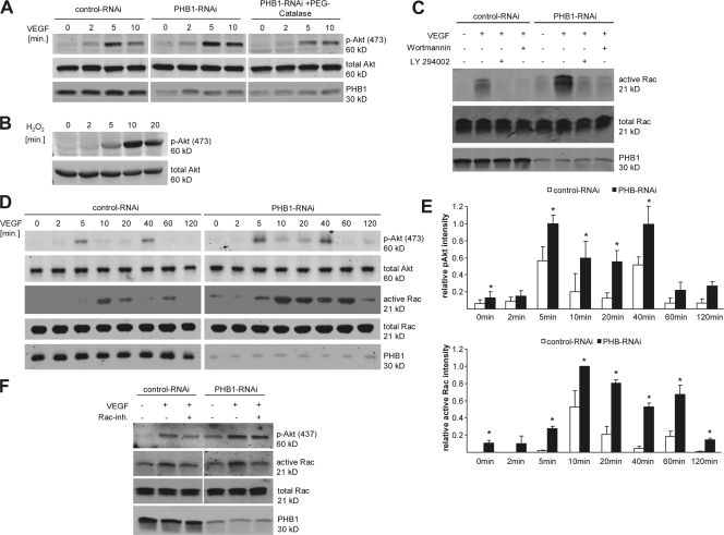
Figure 4.
PHB1 regulates Akt and Rac1 activity. (A) The level of phosphorylated (P473) and total Akt (after 100-ng/ml VEGF stimulation for different time points) was examined via Western blotting after PHB1 knockdown in ECs (middle) in comparison to control cells (left) and PHB1 knockdown cells preincubated with PEG-catalase (right). (B) H2O2 activates Akt phosphorylation, as shown by Western blotting. (C) Rac1 activity assays were performed using control and PHB1 knockdown cells, and active Rac1 pulldowns as well as total cell lysates were Western blotted against Rac1 and PHB1. Cells were treated with 100 ng/ml VEGF and/or PI3-K inhibitors wortmannin (50 nM) and LY294002 (30 μM) as indicated. (D) Phospho-Akt (P473) and active Rac1 were monitored in control and PHB1 knockdown cells upon 100-ng/ml VEGF stimulation for the indicated time points. (E) Quantification of Akt phosphorylation (top) and active Rac1 (bottom) normalized to total Akt and Rac, respectively, from three independent experiments. *, P < 0.05. (F) Rac1 activity in ECs pretreated with VEGF and/or a Rac1 inhibitor as indicated by Western blot analysis of total cell lysates for p-Akt, total Rac1, PHB1, and active Rac1. Error bars show the calculated standard deviation.

