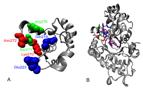
Figure 3.
Predicted coenzyme locations binding sites in PsXR. A) Predicted locations in PsXR for residues that were chosen for CASTing. B) Predicted binding sites of wild-type NT-XR with NADPH. NADPH was in purple. The figures were prepared with VMD 1.8.4 [30], based on a structural model for PsXR obtained via homology modeling using the TASSER-Lite [15, 16, 31]. Red, green and blue colors indicate rounds 1, 2, 3 residues, respectively.

