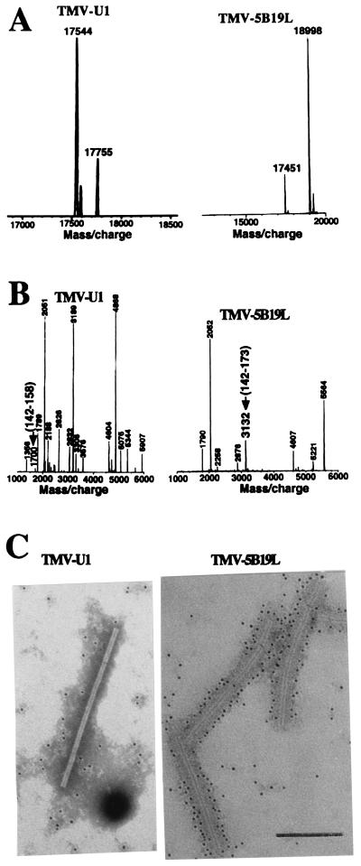
Figure 3.
(A and B) MS analysis of w.t. CP (Left) and CP-5B19L (Right) purified from virus particles. Molecular mass of the CPs untreated (A) or treated with trypsin (B) was determined by matrix-assisted laser absorption/desorption ionization MS. (B) The mass and the amino acid coordinates (between parentheses) of the fragment containing the 5B19L peptide and its homologue in w.t. CP are indicated by arrows. (C) Electron microscopy and immunogold labeling of TMV-5B19L and TMV-U1. Purified virus was subjected to treatment with a monoclonal antibody against the 5B19 peptide followed by goat anti-mouse antibody conjugated to 5-nm gold particles. Samples were negatively stained and analyzed and viewed at 39,000×. (Bar = 200 nm.)

