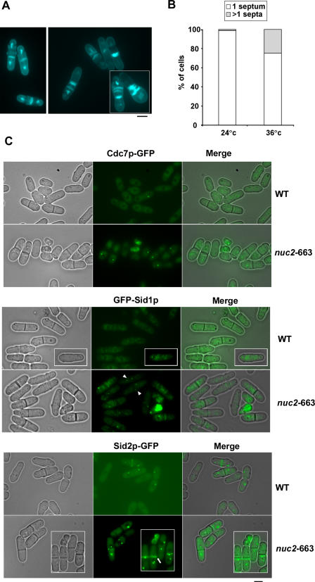
Figure 1. Hyperactivation of SIN Signaling in nuc2-663 Mutant.
(A) nuc2-663 cells were stained with DAPI and aniline blue to visualize nuclei and septum material, respectively. The left panel shows nuc2-663 cells at permissive temperature. The right panel shows nuc2-663 cells at restrictive temperature.
(B) Quantification of multiseptated cells in the nuc2-663 mutant.
(C) Wild-type and nuc2-663 cells expressing GFP tagged versions of Cdc7p, Sid1p, and Sid2p were grown at 36 °C for 4 h and were visualized using fluorescence microscopy. Arrowheads indicate mitotic cells with Sid1p at both SPBs. Arrows indicate cells in which Sid2p-GFP signal persisted at the division site after completion of cytokinesis. Scale bar, 5 μm.

