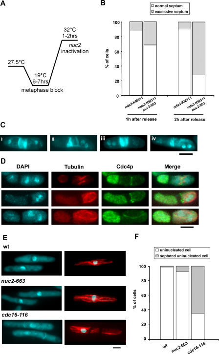
Figure 2. Ectopic Actomyosin Ring and Septum Formation in the nuc2-663 Mutant After Septation.
(A) The diagram schematically illustrates the experimental design.
(B) Quantification of cells with normal or excessive septa after 1–2 h release from metaphase block. At least 200 cells were counted in each category.
(C) Examples of septum patterns in nuc2-663 nda3-KM311 cells. Four examples of cells with excessive septum material are shown. i and ii, multiseptated cells; iii, excessive deposition of septum material at division site; iv, cell with ectopic misoriented septum.
(D) Visualization of actomyosin rings in nuc2-663 nda3-KM311 cells by immunofluorescence microscopy. Top, middle, and bottom panels show cells with actomyosin rings that were formed straight, close to, or misoriented with respect to the previous division site, respectively. Microtubules were stained with TAT-1 antibody, and the actomyosin ring was stained with anti-Cdc4p antibody.
(E) Septum assembly in S-phase–arrested cells. Wild-type, nuc2-663, and cdc16-116 cells arrested in S phase by hydroxyurea treatment were either formaldehyde fixed for DAPI and aniline blue staining or methanol fixed for immunostaining with TAT-1.
(F) Quantification of septated cells versus non-septated cells upon hydroxyurea treatment. At least 300 cells were counted for each category. Scale bar, 5 μm.

