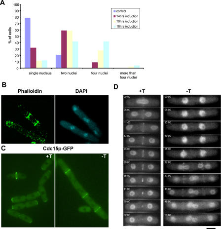
Figure 3. Actomyosin Rings Are Assembled, but Not Maintained, at the Division Site in Cells Overexpressing Nuc2p.
(A) Quantification of the number(s) of nuclei/cell and the frequency of their appearance upon overexpression of Nuc2p. Cells expressing Uch2p-GFP as a nuclear marker were used in the experiment.
(B) Visualization of F-actin. Cells overexpressing Nuc2p were fixed and stained with phalloidin and DAPI to visualize the F-actin cytoskeleton and nuclei, respectively.
(C) Visualization of Cdc15p. Cells carrying Cdc15p-GFP were induced to overexpress Nuc2p and visualized by fluorescence microscopy. +T/−T indicates medium supplemented with or without thiamine.
(D) Time-lapse fluorescence microscopy to image the dynamics of actomyosin ring assembly and constriction. Cells were grown in medium with or without thiamine and imaged by time-lapse microscopy. The nucleus and actomyosin ring were marked by Uch2p-GFP and Rlc1p-GFP, respectively. Scale bar, 5 μm.

