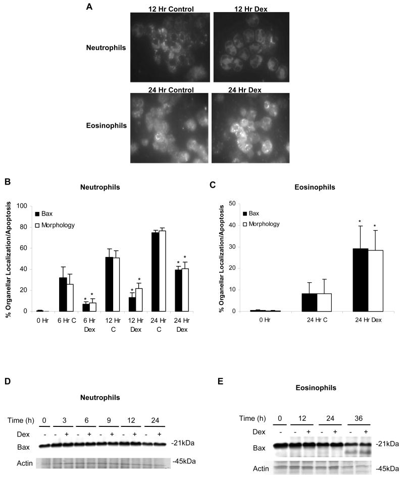
Fig 1.
Organellar, clustered localization of Bax closely corresponds with the induction of apoptosis in neutrophils and eosinophils. (A) Neutrophils and eosinophils were incubated in media alone or with Dex (20 μM). At designated time points, cells were fixed, permeabilized, probed with an anti-Bax antibody, and labeled with an Alexa Fluor 488-conjugated secondary antibody. To compare the movement of Bax with the induction of apoptosis, the percent of neutrophils (B) and eosinophils (C) demonstrating apoptotic morphology was graphed along with the percentage of cells showing clustered labeling of Bax. To examine apoptotic morphology, 1 × 10⁵ cells were collected at each time point, cytocentrifuged, stained with Diff-Quik, and analyzed for apoptotic features including decreased cell volume and condensed nuclei. At least 200 cells were examined per condition. * denotes p<0.05 for Dex-treated cells versus control cells at each time point. At the time points indicated, 1 × 10⁶ neutrophils (D) or eosinophils (E) were harvested and examined for Bax protein expression by Western blot.

