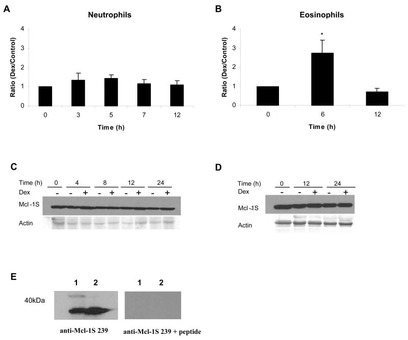
Fig 4.
Mcl-1S protein expression remained constant during Dex treatment in neutrophils and eosinophils, despite changes in mRNA. A) Total RNA was isolated from neutrophils incubated in media alone or with Dex (20 μM) at 0, 3, 5, 7, and 12 h after incubation. B) RNA was isolated from untreated and Dex-treated eosinophils at 0, 6, and 12 h after incubation. Mcl-1S mRNA expression was analyzed by real time PCR and was normalized against GAPDH mRNA expression. The ratio of expression of Mcl-1S mRNA in Dex-treated cells versus control cells was calculated at each time point. * denotes p<0.05 for Dex-treated cells versus control cells at each time point. 1 × 10⁷ neutrophils (C) and 1 × 10⁶ eosinophils (D) were processed and examined for Mcl-1S protein expression by Western blot. E) The specificity of the Mcl-1S 239 antibody was tested by probing blots that contained boiled cell lysates of either 1 × 10⁶ or 1 × 10⁷ total neutrophils. The two sets of samples were run on the same gel, transferred, and then cut and blotted with either the Mcl-1S 239 antibody or the Mcl-1S 239 antibody pre-incubated with the peptide used to raise it (RGPRRWHQECAAGFC, corresponding to the unique amino acids 239-253 of Mcl-1S). The detection of a single band with the antibody alone, and the disappearance of the band upon addition of the peptide confirmed the specificity of the Mcl-1S 239 antibody (representative experiment shown, n=2).

