Figure 1.
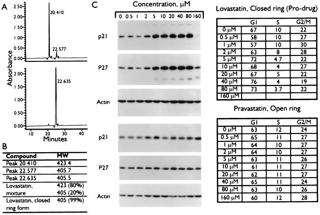
Induction of CKIs by the β-lactone form of lovastatin. (A) Chromatographic separation of lovastatin mixture (Upper) and closed-ring form (Lower) by HPLC analysis as described (15, 25). (B) Fractions corresponding to each HPLC peak were collected and subjected to mass determination by electrospray ionization quadrupole mass spectrometry analysis as described (26). The lovastatin mixture and closed-ring forms also were subjected to mass spectrometry analysis to determine the components of each reagent. (C) MDA-MB-157 tumor cells were treated with the indicated concentrations of lovastatin pro-drug or pravastatin for 36 hr. Cells were harvested and subjected to either flow cytometry (Right) or Western blot analysis with the indicated antibodies (Left).

