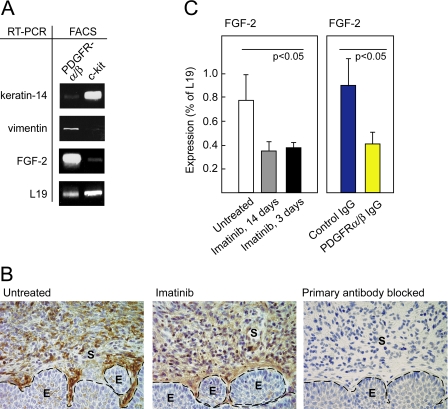
Figure 5. FGF-2 Is Expressed by CAFs and Repressed by Specific Inhibition of PDGF Receptor Signaling.
(A) Analysis of cells isolated by FACS from the cervixes of 3.5-mo-old HPV/E2 mice by sorting for expression of PDGFR-α and -β or c-kit. RT-PCR was performed to assess the expression of FGF-2, the squamous epithelial marker K14, the fibroblast cell marker vimentin, and the housekeeping gene L19 as a loading control.
(B) Representative immunohistochemical staining of FGF-2 in the transformation zone of the uterine cervixes of HPV/E2 mice that had or had not been treated with imatinib displaying CIN3 lesions. The expression pattern was analyzed in at least five different mice of similar histological stage from each treatment group. Parameters: 400× magnification; dotted line marks epithelium-stroma boundary. As a control for specificity, the primary antibody was pre-blocked by incubation with recombinant mouse FGF-2 . E, epithelium; S, stroma.
(C) Expression of FGF-2 analyzed by quantitative RT-PCR following a long (14 d) and a short (3 d) treatment with imatinib (Student t-test, t = 3.5, p < 0.05). Expression of FGF-2 analyzed by quantitative RT-PCR following a 3-d treatment with control IgG or inhibitory antibodies against PDGFR-α and PDGFR-β (Student t-test, t = 3.1, p < 0.05). Note that for technical reasons, a different primer set was used in this experiment compared to the experiment shown in Figure 4C, yielding different absolute values of expression (for details, see Materials and Methods). Error bars indicate the standard error of the mean.

