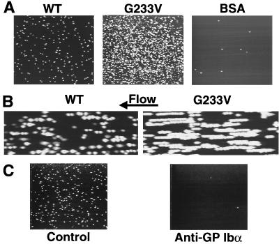
Figure 3.
(A) Images of latex beads coated with GP Ibα amino-terminal domain fragment adhering to immobilized vWF. Fluorescent beads were coated with a 100-μg/ml solution of GP Ibα fragment, which was either normal (WT) or mutant (G233V). Control beads were coated with the same concentration of BSA. Each image, representing a surface area of 1.28 mm2, is a single frame taken from a recording of beads adherent to vWF in a flow field with a wall shear rate of 1,300 s−1. The beads (7,000 per μl) were flowing in a suspension of washed human red cells with hematocrit of 45%. Interactions on the surface were visualized by epifluorescence video microscopy and recorded in real time (30 frames per s). (B) Visual rendition of the translocation (rolling) of beads interacting with immobilized vWF. The beads used in these experiments were coated with solutions of normal or mutant GP Ibα fragment at a concentration of 20 μg/ml. Such a coating concentration resulted in a number of adherent beads that facilitated evaluation of individual interactions. Each image represents a surface area of 0.088 mm2, and 15 consecutive frames, sampled at 1 frame per s from a real-time videotape recording, were digitized and superimposed to show the progressive change in position of the beads on the surface. Each track formed by closely spaced fluorescent dots represents a single bead moving along the direction of flow. (C) Inhibitory effect of a specific monoclonal antibody on the interaction between beads coated with native GP Ibα fragment and immobilized vWF. Experimental conditions as described for A except that, when indicated, the anti-GP Ibα antibody LJ-Ib1 (150 μg/ml) was added to the suspension containing the beads and incubated for 15 min at 22–25°C before perfusion.

