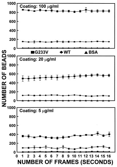
Figure 5.
Quantitative evaluation of the interaction between latex beads coated with GP Ibα amino-terminal domain fragments and immobilized vWF. The beads used in these experiments were coated with solutions at three different concentrations of normal (WT) or mutant (G233V) GP Ibα fragment. Control beads were prepared with equivalent concentrations of BSA. The beads (7,000 per μl) were perfused in suspension with washed human red cells (hematocrit of 45%) at a wall shear rate of 1,300 s−1. The number of beads interacting with immobilized vWF on a surface area of 1.28 mm2 was evaluated in 16 consecutive individual frames from a videotape recording sampled at 1-s intervals. Thus, each frame provided information on surface coverage every second. Two nonoverlapping series of frames obtained between 2 and 5 min after the beginning of perfusion were evaluated for each experimental condition, and the corresponding average values are shown here (±SEM).

