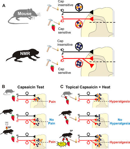
Figure 8. Model Showing the Patterns of C-fiber Innervation in the Spinal Cord of Mouse and Naked Mole-Rat.
(A) Both mouse and naked mole-rat have populations of capsaicin-insensitive and capsaicin-sensitive C-fiber nociceptors. Symbols (hammer, match, chilli pepper) indicate the type of stimuli (mechanical, thermal, chemical irritant) that can generate spiking activity in each population. In both species, capsaicin-insensitive and capsaicin-sensitive fibers terminate in the superficial lamina of the dorsal horn (above the dashed line in the spinal cord schematic). A major difference between species is that in the naked mole-rat, a substantial number of capsaicin-sensitive fibers also provide synaptic input to deep lamina. Another major difference is that C-fibers in naked mole-rats lack the neuropeptides SP and CGRP (yellow dots represent neuropeptides, blue dots represent glutamate).
(B) Capsaicin test: Injection of capsaicin into the skin of the paw induces pain behaviors (licking) in mice but not naked mole-rats, except in naked mole-rats that have received an intrathecal infusion of SP.
(C) Topical capsaicin + heat: Topical application of capsaicin induced thermal hyperalgesia in mice but not naked mole-rats, except in naked mole-rats that have received gene therapy with the PPT gene.

