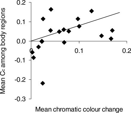
Figure 4. Relationship between Colour Change and Colour Contrast among Body Regions.
The plot shows the average colour change for all three body regions (top, mid-, and bottom flanks) regressed against the mean chromatic contrast (CC) of dominant signals among adjacent body regions (r 2 = 0.18, p = 0.04, with one outlier removed). The regression is based on Felsenstein's independent contrasts (FIC, positivized on the x-axis), regressed through the origin, with the regression slope indicated by the line. There are N – 1 contrasts and one outlier was removed, resulting in 19 points. As in Figure 3, the outlier is the contrast between B. pumilum from Stellenbosch and B. pumilum from Vogelgat.

