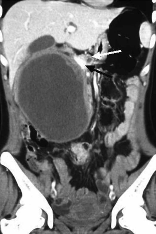
Figure 1. .
Coronal reformation of contrast-enhanced abdominal MDCT showing a large cystic mass with well defined smooth walls. Also seen is an irregular solid component along the left lateral and superior walls. The mass is in close contact with the pancreatic head (white arrow) and the portal vein is seen medial to it (black arrow).

