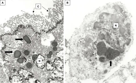
Figure 4. TEM of formalin-fixed, paraffin-embedded lung from KD patient 1.
A, ciliated bronchial epithelium demonstrating electron-dense apical ICI (block arrows). B, alveolar macrophage, demonstrating perinuclear, finely granular spheroid bodies similar to those seen within the bronchial epithelium (block arrow). N = nucleus, C = cilia. A = 9,500X, B = 26,000X.

