Figure 2.
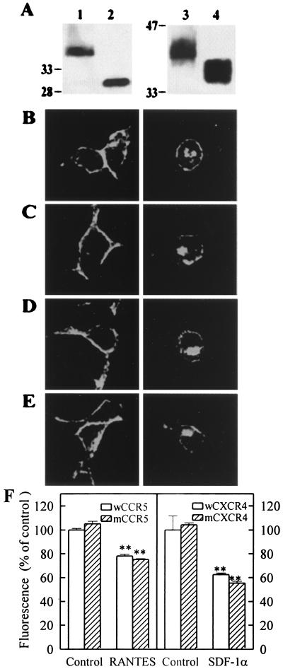
Expression and internalization of the five-TM chemokine receptors. (A) The expression of wCCR5 (lane 1), mCCR5 (lane 2), wCXCR4 (lane 3), and mCXCR4 (lane 4) in transiently transfected HEK293 cells was detected by immunoprecipitation and Western blotting with 12CA5. (B–E) Cells transiently transfected with wCCR5 (B), mCCR5 (C), wCXCR4 (D), and mCXCR4 (E) were incubated without (Left) or with (Right) 10 nM agonist (RANTES for CCR5 and SDF-1 for CXCR4) for 30 min, and internalization of receptors from the cell surface was analyzed by laser confocal fluorescence microscopy using 12CA5 and FITC-conjugated anti-mouse IgG. (F) Similarly, internalization of the wild-type and five-TM mutant CCR5 or CXCR also was determined by using flow cytometry after incubation with or without (control) 10 nM chemokine. Untransfected cells or mock-transfected cells showed negative staining under the same conditions (not shown). Pictures shown in A–E are representative of two separate experiments. The data in F indicate averages and error ranges of two independent determinations in duplicate. ∗∗, P < 0.01 compared with unpretreated controls.

